Every day with Thyroid Eye Disease raises a new question

A free app for everything Thyroid Eye Disease asks of you, grounded in medical science and the experience of others, so you don't have to figure it out alone.

One app for everything Thyroid Eye Disease asks of you
Personalized answers before, after, and in between every appointment.


What to know about Thyroid Eye Disease
The basics on causes, types, symptoms, treatment and diagnosis, written from medical guidelines and reviewed by our in-house doctors. Open the parts that apply to you.
What is thyroid eye disease?
Thyroid eye disease (TED), also called Graves' orbitopathy, is an autoimmune inflammatory condition affecting the tissues and muscles around the eyes. It is most often associated with Graves' disease but can occur in people with normal or low thyroid hormone levels.
TED usually has an active inflammatory phase followed by a more stable inactive phase. Eye activity and severity do not always parallel thyroid blood-test results. mama health's guide to what thyroid eye disease is covers the condition in full.
What causes thyroid eye disease?
Thyroid eye disease occurs when immune reactions target proteins shared by thyroid tissue and cells within the eye socket. Inflammation causes swelling of orbital fat and eye muscles, which can push the eyes forward and restrict movement.
Smoking is the strongest risk factor that can be changed, and it also affects how well treatment works, so support to stop is part of TED care rather than something to manage alone. Unstable thyroid hormone levels, radioactive iodine in selected higher-risk situations, older age, diabetes and certain antibody patterns may also influence severity. Removing the thyroid is sometimes discussed in this context, covered in thyroid removal and thyroid eye disease.
Treatment options
Mild TED may be managed with lubricating drops, gels, head elevation, prism lenses, stopping smoking and correction of thyroid dysfunction. Active moderate-to-severe TED may require corticosteroids, targeted biologic therapy or other immune-modulating treatment. mama health's guide to thyroid eye disease treatment options goes through each.
Surgery is generally considered after inflammation has stabilized and may include orbital decompression, eye-muscle surgery and eyelid surgery. Urgent treatment is required when the optic nerve is compressed or the cornea is at risk. Whether any of this resolves the condition is a common question, covered in can thyroid eye disease be cured. Questions between appointments can be put through mama health, which is free thyroid eye disease support grounded in medical science and the experience of others.
Living with thyroid eye disease
Keeping thyroid hormone levels stable and attending regular ophthalmology assessments during the active phase are the two things that matter most. Sunglasses, lubricants, sleeping with the head elevated and taping the eyelids closed at night under professional guidance may reduce exposure symptoms.
Double vision and changes around the eyes can affect driving, work and emotional wellbeing. Temporary prisms, occupational adjustments and psychological support may all be useful while activity is changing. mama health's guides to managing double vision and what people living with TED have found helps cover the practical side, and real-life challenges with TED covers what other people describe. What changes week to week can be kept in one place in the mama health app, a free thyroid eye disease app built with in-house doctors.
What happens after the active phase?
Many cases remain mild, while others leave lasting changes in eye position, the eyelids or vision. Controlling risk factors early and treating active inflammation appropriately reduces complications, and staged reconstructive surgery after TED becomes inactive can improve both function and how the eyes look.
Who is at risk of developing thyroid eye disease?
Risk factors include Graves' disease, smoking or secondhand smoke exposure, high thyroid-receptor antibody levels, poorly controlled thyroid dysfunction, older age, diabetes and previous thyroid eye disease.
Complications include corneal damage, persistent double vision, elevated eye pressure, lasting changes to the eyes and eyelids, and compressive optic neuropathy, which can cause permanent sight loss. mama health's guide to how patients experience thyroid eye disease covers what that is like in practice.
What are the symptoms of thyroid eye disease?
Symptoms include dry, gritty, watery, red or painful eyes; sensitivity to light; eyelid swelling or retraction; a staring appearance; bulging eyes; pressure behind the eyes; pain with eye movement; blurred or double vision; reduced eye movement; and difficulty closing the eyelids. More severe TED may reduce sharpness of vision, color vision or the field of vision. mama health's guide to thyroid eye disease symptoms covers each in more detail.
Bulging is the symptom most people have heard of, and it does not affect everyone, which do the eyes always bulge with thyroid eye disease explains.
How is thyroid eye disease diagnosed?
An ophthalmic examination assesses visual acuity, pupils, color vision, visual fields, eye pressure, corneal exposure, eyelid position, proptosis and eye movements. Clinical activity scores help distinguish active inflammation from stable residual changes.
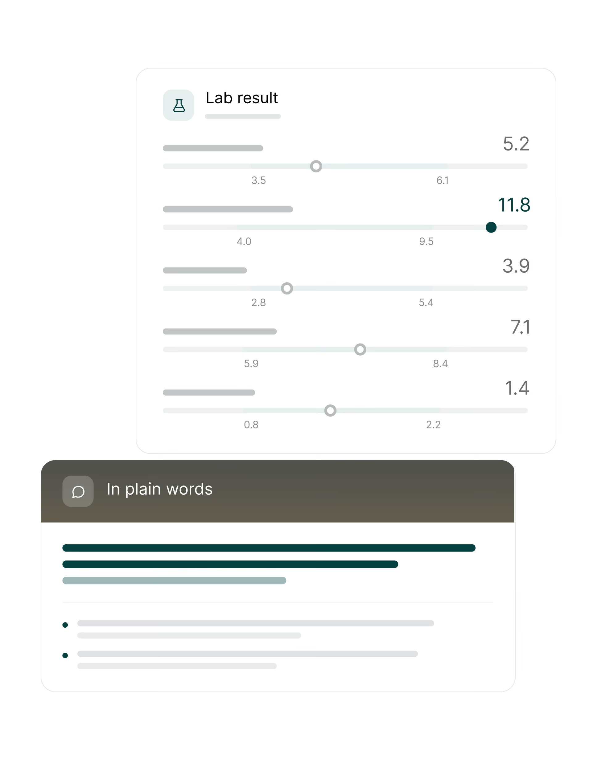
Blood tests measure thyroid function and thyroid-receptor antibodies, and mama health's guide to thyroid ultrasound covers one of the tests used alongside them. Orbital CT or MRI evaluates enlarged eye muscles, crowding around the optic nerve, inflammation or alternative causes. Optical coherence tomography or visual-field testing may be used when optic nerve involvement is suspected. Results and letters can be read back in plain language in the mama health app, a free thyroid eye disease support app.
What stands behind mama health
Medical oversight, safeguards around your data, and a clear purpose for every anonymized experience shared.
.avif)







Reviewed by our doctors
Our doctors review the medical sources behind every AI answer. Designed for high accuracy from your first chat.

Anonymized for good
Your personal data is encrypted and never shared. We turn it into anonymized insights. All under strict European data standards.

Contributing to research
Partners learn from anonymized patient experiences. That helps push research and better treatments forward.
All conditions we add to mama health go through careful checks.



Frequently asked questions
The report will appear in your inbox shortly✅
If you would like to explore how real-time patient insights can strengthen your commercial strategies, book a chat through the link below.